
Translate this page into:
A 70-year-old woman with low back pain

*Corresponding author: Shrishail Adke, Department of Radiology, Deenanath Mangeshkar Hospital, Pune, Maharashtra, India. shri.adke@gmail.com
-
Received: ,
Accepted: ,
How to cite this article: Adke S, Desai S. A 70-year-old woman with low back pain. Indian J Musculoskelet Radiol. 2025;7:139-41. doi: 10.25259/IJMSR_72_2024
PART 1. QUESTION
Clinical history
A 70-year-old lady presented with lower back pain.
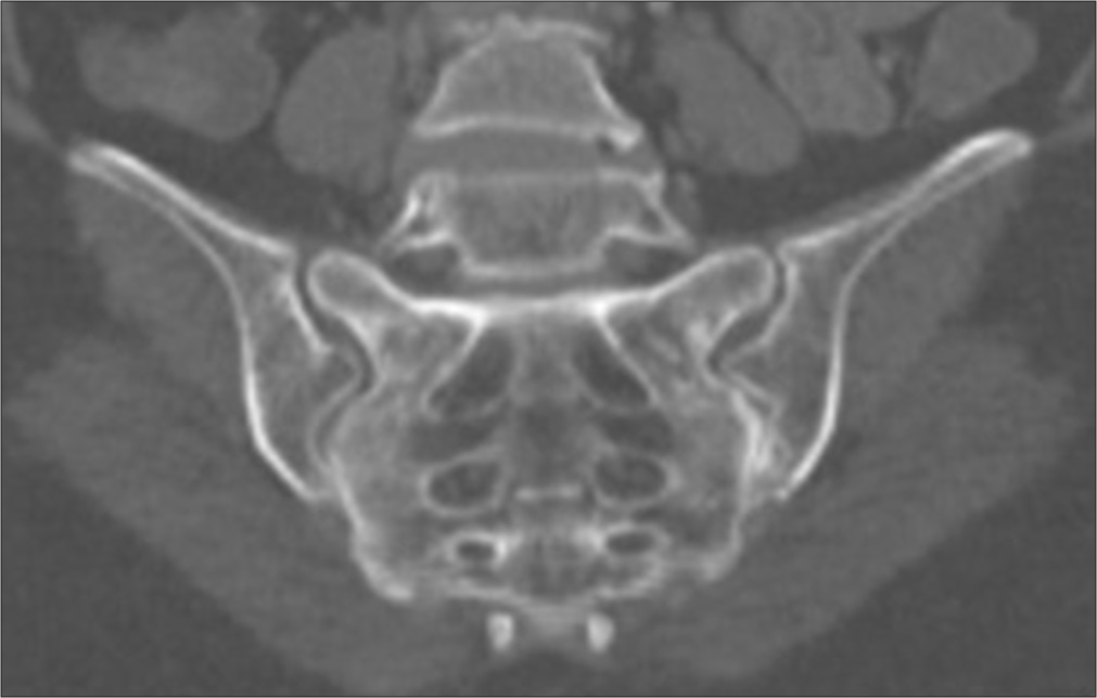
- Oblique coronal computed tomography image of sacroiliac joints.
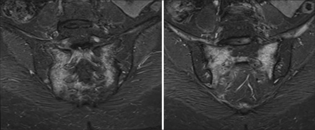
- Sequential oblique coronal short tau inversion recovery (STIR) images of sacroiliac joints.
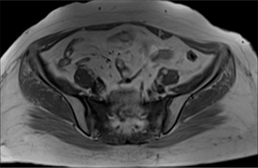
- Axial T1 non-fat suppressed image of sacroiliac joints.
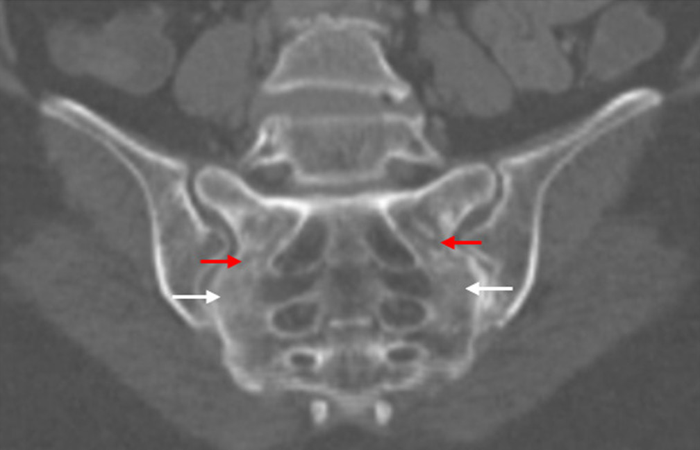
- Oblique coronal computed tomography image of sacroiliac joint reveal ill defined sclerosis in bilateral sacral alae (white arrows) with linear lucencies (red arrows) suggestive of fracture line; however, a breach in the cortex is not evident.
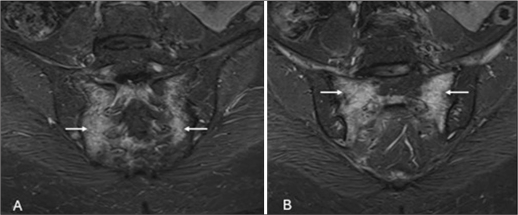
- Sequential oblique coronal short tau inversion recovery images (STIR) images of sacroiliac joints reveal: (white arrows in A) STIR hyperintensity s/o edema in bilateral sacral alae lateral to the neural foramina and medial to the sarcoiliac joints and (arrows in B) hypointense fractures lines parallelly oriented to the sacroiliac joints.
PART 2: ANSWER
Findings
Diagnosis: Bilateral sacral insufficiency fractures.
DISCUSSION
An insufficiency fracture is a subtype of stress fracture that occurs due to physiological stress on weakened bones (commonly due to osteoporosis). They are most frequently observed in elderly women. In the pelvis, common sites for insufficiency fractures include the sacrum and pubic bone.[1] These patients often present with low back pain in the absence of significant trauma. Sacral insufficiency fractures (SIFs) often exhibit a characteristic H-shaped pattern, referred to as the “H-sign” or “Honda sign” on bone scans. This pattern is characterized by vertical fracture lines that run parallel to the sacroiliac joints in the bilateral sacral alae, as well as a horizontal fracture line through the sacral body.[2]
Magnetic resonance imaging (MRI) is considered the most sensitive and specific diagnostic tool for sacral stress fractures. On MRI, the fracture site typically demonstrates marked edema, with the fracture line appearing as a hypointense band or line on both T1 and T2 sequences or as a fluid-filled cleft.[3] In contrast, computed tomography (CT) is less sensitive than MRI or nuclear imaging and may fail to detect fractures if cortical continuity is maintained.[4]
Most sacral insufficiency fractures respond well to conservative management, with surgical intervention rarely required.[5]
Looser zones also known as Milkman lines are type of insufficiency fractures and commonly involve pubic ramus, medial femoral neck, medial proximal femoral shaft (weight-bearing sites) and scapula, iliac wing, ulna, ribs (non-weight-bearing sites). In weight-bearing bones, they occur in the same locations as insufficiency fractures and, therefore, are often diagnosed and treated as fractures.[6] Hence, entire pelvis should be evaluated in elderly patients with low back ache.
DIFFERENTIALS
Marrow infiltrative neoplasm: diffuse and varied involvement of other bones with abnormal marrow signal intensity.
Sacroiliitis: predominantly peri-articular involvement of sacrum and iliac bones with articular surface changes.
Acknowledgment
We acknowledge the help extended by Dr. Stanzin Spalkit in editing the manuscript.
Ethical approval
Institutional Review Board approval is not required.
Declaration of patient consent
Patient’s consent not required as patients identity is not disclosed or compromised.
Conflicts of interest
There are no conflicts of interest.
Use of artificial intelligence (AI)-assisted technology for manuscript preparation
The authors confirm that there was no use of artificial intelligence (AI)-assisted technology for assisting in the writing or editing of the manuscript and no images were manipulated using AI.
Financial support and sponsorship: Nil.
References
- Pelvic insufficiency fracture in severe osteoporosis patient. Hip Pelvis. 2017;29:120-6.
- [CrossRef] [PubMed] [Google Scholar]
- Honda sign and variants in patients suspected of having a sacral insufficiency fracture. Clin Nucl Med. 2005;30:165-9.
- [CrossRef] [PubMed] [Google Scholar]
- Superiority of MRI for evaluation of sacral insufficiency fracture. J Clin Med. 2022;11:4968.
- [CrossRef] [PubMed] [Google Scholar]
- Imaging of insufficiency fractures. Eur J Radiol. 2009;71:398-405.
- [CrossRef] [PubMed] [Google Scholar]
- Imaging and treatment of sacral insufficiency fractures. AJNR Am J Neuroradiol. 2010;31:201-10.
- [CrossRef] [PubMed] [Google Scholar]
- A mechanism of the production of pseudofractures in osteomalacia (Milkman's syndrome) Radiology. 1954;62:388-95.
- [CrossRef] [PubMed] [Google Scholar]


