Translate this page into:
Displaced flap tear of the triangular fibrocartilage complex – A case report

*Corresponding author: Karan Asthana, Department of Radiology, Deenanath Mangeshkar Hospital, Pune, Maharashtra, India. karanasthana12@gmail.com
-
Received: ,
Accepted: ,
How to cite this article: Asthana K, Desai S. Displaced flap tear of the triangular fibrocartilage complex – A case report. Indian J Musculoskelet Radiol. 2024;6:153-5. doi: 10.25259/IJMSR_43_2024
Abstract
Triangular fibrocartilage complex (TFCC) tears are common after acute wrist injuries and have been classified. However, there are complex patterns of TFCC injuries which can sometimes be encountered and are not mentioned in standard TFCC injury classification. Flap tears are very rare pattern of TFCC injury (0.5%) and post a diagnostic and management challenge. Magnetic resonance imaging is the investigation of choice in such cases. This case represents a unique pattern of TFCC injury in which the central disc was displaced in the volar aspect of distal radioulnar joint space.
Keywords
Distal radioulnar joint
Displaced
Flap
Triangular fibrocartilage complex
Tear
INTRODUCTION
Post-traumatic injuries to the triangular fibrocartilage complex (TFCC) are frequently encountered in day to day practice. Palmer classification of traumatic TFCC tears is well documented. However, additional tear of the TFCC has been described in literature having an incidence of 0.5%.[1] These do not have a well-documented classification system. In this case report, we describe a complex TFCC tear in which a fragment of the central disc was displaced into the distal radioulnar joint (DRUJ) space.
CASE REPORT
A 30-year-old male patient presented with pain in the left wrist joint following trauma. On clinical examination, tenderness was present in the ulnar aspect of the wrist. There was pain during pronation – supination movement. Features of DRUJ instability were present as evidenced by positive piano key sign.
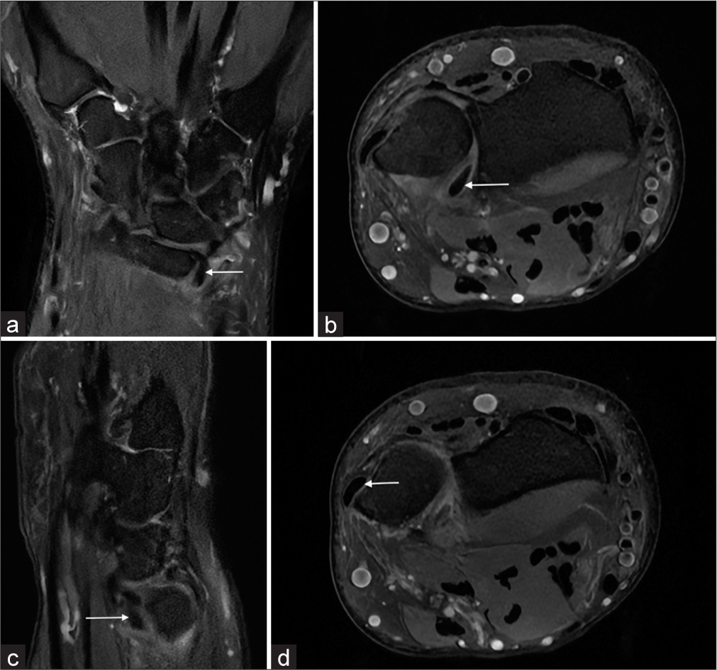
Magnetic resonance imaging (MRI) of the patient was performed. MRI revealed displacement of the central disc of the triangular fibrocartilage (TFC) into the DRUJ space. Fluid was seen within the distal ulnar radioulnar joint space.
There was medial subluxation of the extensor carpi ulnaris tendon.
DISCUSSION
A TFCC is formed by ligaments, cartilage, and tendon. The parts include triangular fibrocartilaginous articular disc, volar and dorsal radioulnar ligaments, volar ulnotriquetral and ulnolunate ligaments, ulnar collateral ligament, extensor carpi ulnaris tendon sheath, and ulnar meniscal homolog.[2]
TFCC injuries often occur after a patient sustains an axial load on a hyperpronated wrist.[3]
With the wrist in pronation, the relative dorsally displaced ulnar head may be reduced volarly to the radius with direct pressure or elicit pain, indicating an abnormal piano key sign.[4]
Displaced TFCC tear can cause pain along with mechanical symptoms that warrant flap excision.[3] The displaced flap tears are most commonly located in the volar recess of the DRUJ.[3]
MRI is the investigation of choice for assessment of TFCC tear.
Displaced TFCC tear can be missed at surgery, surgeons hence advocate for pre-operative MRI for diagnosis of flap tears displaced into the radioulnar joint.[5]
The coronal proton density fat-suppressed image of the left wrist joint revealed a displaced fragment of the central disc of the TFC in the volar aspect of the DRUJ given the appearance of “comma sign”[1] [Figure 1a].

- (a) The coronal proton density fat-suppressed image of the left wrist join reveals a dispalced fragment of the central disc of the triangular fibrocartilage (TFC) in the volar aspect of the distal radioulnar joint (arrow) given the appearance of the “comma sign.” (b) Axial proton density fat-suppreseed image of the left wrist joint reveals a flipped fragment of the TFC (central disc) in the volar aspect of the distal radioulnar joint (arrow). (c) The sagittal proton density fat-suppressed image of the left wrist joint reveals a displaced fragment of the TFC, central disc displaced in the volar aspect of the distal radioulnar joint (arrow) with surrounding edema. (d) Axial proton density fat-suppressed image of the left wrist joint shows medial subluxation of the extensor carpi ulnaris tendon (arrow).
Axial proton density fat-suppressed images of the left wrist joint revealed a flipped fragment of the TFC (central disc) in the volar aspect of the DRUJ [Figure 1b].
The sagittal proton density fat-suppressed images of the left wrist joint revealed a displaced fragment of the TFC, central disc displaced in the volar aspect of the DRUJ with surrounding soft-tissue edema [Figure 1c].
Axial proton density fat-suppressed images of the left wrist joint showed medial subluxation of the extensor carpi ulnaris tendon [Figure 1d].
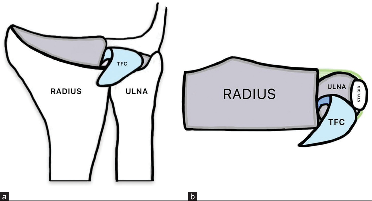
Figures 2a and 2b represent simplified coronal and axial diagrammatic representation of the index case showing the displaced fragment of the TFC (in blue).

- (a) Simplified coronal diagrammatic representation of the index case showing the displaced fragment of the triangular fibrocartilage (TFC) (in blue). (b) Simplified axial diagrammatic representation of the index case showing the displaced fragment of the TFC (in blue).
CONCLUSION
Complex tear of the TFC does not have a well-documented classification system and is a diagnostic and management challenge. Displaced tears of the central disc lying in the dorsal radioulnar joint are not seen on arthroscopy. Pre-operative diagnosis is necessary for a better surgical outcome and MRI is the investigation of choice. Radiologists should be well versed with the displaced flap tears of the TFC. A defect in the normal bulk of TFCC along with a displaced fragment in the joint recess should raise suspicion of this condition. Often on MRI, a displaced flap pedicle can be visualized and connected to the native TFC.
Ethical approval
Institutional Review Board approval is not required.
Declaration of patient consent
Patient’s consent not required as patients identity is not disclosed or compromised.
Conflicts of interest
There are no conflicts of interest.
Use of artificial intelligence (AI)-assisted technology for manuscript preparation
The authors confirm that there was no use of artificial intelligence (AI)-assisted technology for assisting in the writing or editing of the manuscript and no images were manipulated using AI.
Financial support and sponsorship
Nil.
References
- Displaced flap tears of the triangular fibrocartilage complex: Frequency, flap location, and the “Comma” sign on wrist MRI. AJR Am J Roentgenol. 2021;217:707-8.
- [CrossRef] [PubMed] [Google Scholar]
- It is not only the meniscus that flips-a case of TFCC tear with a flipped fragment in the distal radioulnar joint. Indian J Radiol Imaging. 2023;33:129-31.
- [CrossRef] [PubMed] [Google Scholar]
- Surgical repair of acute TFCC injury. Hand (NY). 2020;15:674-8.
- [CrossRef] [PubMed] [Google Scholar]
- MRI findings in bucket-handle tears of the triangular fibrocartilage complex. Skeletal Radiol. 2018;47:419-24.
- [CrossRef] [PubMed] [Google Scholar]


