
Translate this page into:
Extradigital Glomus Tumor on Hypothenar Region Diagnosed on Ultrasound – A Case Report

-
Received: ,
Accepted: ,
How to cite this article: Kachare M, Khan A, Joshi U, Patil S. Extradigital Glomus Tumor on Hypothenar Region Diagnosed on Ultrasound – A Case Report. Indian J Musculoskelet Radiol 2021;3(1):60-3.
Abstract
We report a case of 32-year-old female with a 6 months history of excruciating pain in hypothenar region of the left hand. Pain was aggravated by pressure, touch, and cold temperature. Musculoskeletal ultrasonography revealed – a well-defined, hypoechoic lesion in deep dermis, and subcutaneous fat in the left hypothenar eminence with mixed arterial and venous signals within on Doppler study, suggestive of – subcutaneous vascular lesion and diagnosis of glomus tumor was suggested. The patient underwent excision of the lesion. Pathological examination of the specimen showed a glomus tumor and excluded malignant transformation to glomangiosarcoma. Extradigital glomus tumor can be diagnosed on ultrasound with high confidence in appropriate clinical setting.
Keywords
Glomus tumor
Ultrasonography
Subcutaneous fat
Dermis
Hypothenar

INTRODUCTION
Glomus tumors are benign mesenchymal tumors of neuromyoarterial glomus bodies and are most commonly found in the subungual area of the fingers. However, extradigital glomus tumors are rare and are difficult to diagnose due to their low incidence and non-specific symptoms and usually missed during the physical examination. On ultrasonography (USG), extradigital glomus tumor (EDGT) typically appears as a well-defined, small, hypoechoic, homogeneous, or slightly inhomogeneous nodule which are painful under probe pressure. Intranodular vascularity is always present.
CASE REPORT
A 32-year-old woman presented to orthopedic surgeon with excruciating pain in hypothenar region of the left hand for 6 months. Pain was aggravated by pressure, touch, and cold temperature.
Ultrasound of the hand was done with 17 MHz high frequency transducer in musculoskeletal setting on Toshiba Xario system (Otawara, Tochigi, Japan). On USG, a 2.5 × 2.2 mm well defined, hypoechoic lesion was noted in deep dermis and subcutaneous fat in the left hypothenar eminence. This soft-tissue lesion revealed internal mixed arterial and venous signals on Doppler study. At the site of the USG abnormality, a small bluish lesion was seen in the hypothenar region.
Lesion was excised under local anesthesia by orthopedic surgeon and the specimen was sent for histopathological examination.
The histopathology revealed small circumscribed tumor composed of clusters of uniform round to oval cells with round nuclei and eosinophilic cytoplasm. The cells were perivascular in location with a few capillaries in the centers of clusters. The stroma was scant and there was no evidence of malignancy. These findings were consistent with extradigital glomus tumor.
DISCUSSION
Glomus tumors are slow-growing benign neoplasms of neuromyoarterial glomus bodies origin. These are usually located in the fingers and particularly in the subungual bed. However, EDGTs predominate in extremities, such as the thigh, leg, and forearm.[1]
Extradigital glomus tumors are difficult to diagnose due to their rarity and non-specific symptoms and may be overlooked on physical examination.[2] In this case, the patient presented with pain in the left hypothenar region which was aggravated on pressure and exposure to the variation in temperature [Figure 1]. Pain on temperature variation is thought to be due to reflex vasodilatation or it may be related to contraction of myofilaments.[3]
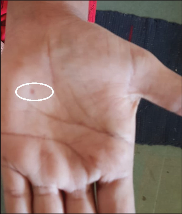
- Picture of hand reveals small bluish cutaneous lesion in hypothenar region.
Glomus tumor is considered to be more frequently described in young and middle-age adults as glomus bodies are absent in children younger than 1 year of age, and they tend to atrophy in the older people.[4]
On USG, the lesion typically appears as a well-defined, hypoechoic, circumscribed oval subcutaneous nodule, with slight posterior acoustic enhancement, and tenderness on probe pressure [Figure 2].[5] Differently from hemangioma EDGT is firm and does not change in shape under probe-mediated pressure.[3]
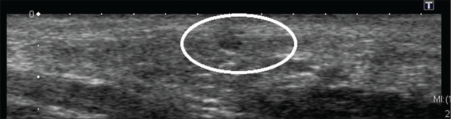
- A well-defined hypoechoic ovoid lesion is seen in deep dermis and subcutaneous fat of the left hypothenar eminence on gray scale ultrasonography.
Although EDGTs are small in size, it reveals several vessels internally. High frequency probe with color Doppler and minimal pressure application allows the demonstration of typical but not constant, hypervascularity [Figure 3]. The presence of a single feeding large artery and single or multiple draining veins is very typical. Spectral Doppler demonstrates both arterial flow and to a relatively lesser degree, venous flow [Figure 4].[3]
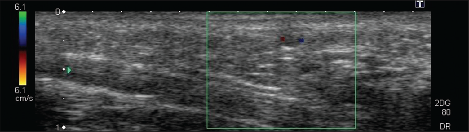
- On color Doppler ultrasonography study, the hypoechoic lesion shows mixed vascularity.
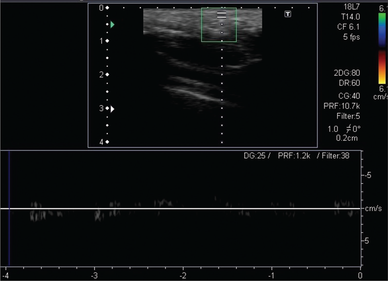
- Low velocity arterial and venous signals are noted in the lesion on the spectral Doppler study.
The imaging or histopathological findings reveal absent features of malignancy [Figure 5]. It is unusual for a glomus tumor to demonstrate atypical or malignant histopathological characteristics but malignant transformation to glomangiosarcoma and metastasizing features such as nuclear atypia, infiltrative growth pattern, and multicentricity although rare have been described. Malignancy can be suspected in the presence of multiple, large, deep, and heterogeneous EDGTs.[6]
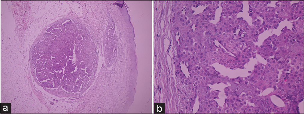
- (a) Histologically on hematoxylin and eosin (H and E) stain, original magnification ×40 – Low-power microscopic view showing tumor as a well-defined small circumscribed lesion in deep dermis and subcutaneous fat layer. (b) H and E stain, original magnification ×100 – Medium-power microscopic view showing the tumor is composed of clusters of uniform round to oval cells with round nuclei and eosinophilic cytoplasm along capillary cluster.
CONCLUSION
An EDGT is a clinically painful lesion with indeterminate signs which can be diagnosed on USG with high frequency transducer. Correlating the clinical presentation with the gray scale sonography and color Doppler findings is helpful to suspect an EDGT. However, histopathology is required for the definitive diagnosis of tumor.
Declaration of patient consent
The authors certify that they have obtained all appropriate patient consent.
Financial support and sponsorship
Nil.
Conflicts of interest
There are no conflicts of interest.
References
- Color Doppler sonography of extradigital glomus tumors. J Ultrasound Med. 2017;36:231-8.
- [CrossRef] [PubMed] [Google Scholar]
- Glomus tumor of the fingertips: A frequently missed diagnosis. J Family Med Prim Care. 2019;8:904-8.
- [CrossRef] [PubMed] [Google Scholar]
- Painful growth on right index finger, Subungual glomus tumor. Dermatol Online J. 2011;17:12.
- [Google Scholar]
- Ultrasound of soft tissue masses of the hand. J Ultrason. 2012;12:381-401.
- [CrossRef] [PubMed] [Google Scholar]
- Malignant glomus tumor of the lung with multiorgan metastases: Case report and literature review. Onco Targets Ther. 2015;8:1909-14.
- [CrossRef] [PubMed] [Google Scholar]


