
Translate this page into:
Imaging findings of thrower’s elbow in a fast bowler: A case report


*Corresponding author: Neeti Ajay Gupta, Department of Radiology, Sant Parmanand Hospital and Delhi Institute of Trauma and Orthopaedics, New Delhi, Delhi, India. neetigpt92@gmail.com
-
Received: ,
Accepted: ,
How to cite this article: Gupta NA, Varshney A. Imaging findings of thrower’s elbow in a fast bowler: A case report. Indian J Musculoskelet Radiol. 2024;6:40-4. doi: 10.25259/IJMSR_61_2023
Abstract
“Thrower’s elbow” is a term that has been used to describe a constellation of imaging findings seen in players involved in overhead throwing sports, with maximum literature found on baseball injuries. On reviewing the literature, no cases of thrower’s elbow have been described in fast bowlers or cricketers. This case report describes a fast bowler with lateral elbow pain secondary to impaction injury and capitellar osteochondral lesion and concomitant findings of chronic overuse injury involving the ulnar collateral ligament, which is a part of the thrower’s elbow.
Keywords
Elbow
Thrower’s elbow
Medial collateral ligament
Osteochondral lesion

INTRODUCTION
Throwing athletes are prone to elbow injuries secondary to high stress and valgus load in both acute and chronic settings.[1] Thrower’s elbow includes a constellation of imaging findings seen in players involved in overhead throwing sports, including baseball, softball, football, and javelin throwing. The condition has not yet been widely described in cricket bowlers. On reviewing the literature, we found a single case report of a typical case of valgus hyperextension syndrome in an international cricket fast bowler.[2] The various elbow injuries seen in throwing athletes are related to excessive valgus load, which may exceed the inherent strength of medial elbow joint stabilizers, resulting in acute and chronic overuse injuries. Imaging evaluation includes radiographs, ultrasound (US), and magnetic resonance imaging (MRI), with a limited role of computed tomography scans in a few cases.
Our case report describes a fast bowler with complaints of elbow pain, showing findings of thrower’s elbow on imaging. On reviewing the literature, no cases of thrower’s elbow have been described in fast bowlers or cricketers.
CASE REPORT
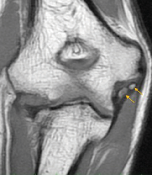
A 32-year-old cricketer (fast bowler) presented with complaints of pain over the lateral aspect of the right elbow and felt more on throwing. There was no significant history of injury or swelling. On examination, he had tenderness over the lateral aspect of the joint with features of valgus instability. There was no significant restriction on the range of movements. Antero-posterior radiograph of the right elbow joint showed heterotopic ossification adjacent to the medial epicondyle of the humerus [Figure 1]. US evaluation of the elbow joint revealed a thickened and heterogeneous ulnar collateral ligament (UCL), with a hypoechoic tear at its humeral attachment and calcific foci within its proximal portion, corresponding to the heterotopic ossification seen on the radiograph [Figure 2]. No abnormality could be seen at the site of lateral elbow pain on radiographs or ultrasound. MRI was thus performed for further evaluation. MRI showed thickening with intra-ligamentous heterotopic ossification involving the proximal portion of the anterior band of the UCL, with a full-thickness tear at its humeral attachment [Figures 3 and 4]. In addition, it also revealed a small early osteochondral lesion (OCD) and subcortical marrow edema involving the posterior aspect of humeral capitellum, possibly secondary to impaction injury, along with thickening and T2-weighted hypointense fibrosis involving the posterolateral joint capsule [Figures 4 and 5a and b]. This constellation of imaging findings of a chronically injured, thickened UCL with calcification, posterior capitellar impaction injury, and posterolateral capsular thickening was attributed to “thrower’s elbow.”

- Anteroposterior radiograph of the right elbow joint showing heterotopic ossification adjacent to the medial epicondyle of humerus (arrow).

- Ultrasound image of the medial elbow in long axis shows a thickened and heterogeneous ulnar collateral ligament, with calcific foci within its proximal portion (arrows).
- Coronal T1-weighted magnetic resonance image shows thickening with intraligamentous heterotopic ossification involving the proximal portion of the anterior band of the ulnar collateral ligament (arrows).

- Coronal PD fat-saturated magnetic resonance image shows thickening of the anterior band of the ulnar collateral ligament, with a full-thickness tear at its humeral attachment (arrow).

- (a) Coronal PD fat-saturated magnetic resonance (MR) image shows subcortical marrow edema (arrow) involving the posterior aspect of humeral capitellum. (b) Sagittal T2-weighted (T2W) MR image also shows thickening and T2W hypointense scarring involving the posterolateral joint capsule (arrow).
DISCUSSION
The elbow joint bears significant stress during the overhead throwing action of athletes participating in sports such as baseball, javelin throwing, and softball. The various elbow injuries seen in them are typically related to excessive valgus load. However, the condition has not yet been widely described in fast bowlers. During the delivery phase of bowling, fast bowlers in cricket are not allowed to flex their elbows more than 15° in their action to deliver the ball.[2] The action of “throwing” or “chucking” is actually deemed to be illegal in cricket. This principally neutralizes the chances of a “thrower’s elbow” happening in them. Contrary to this fact, the authors believe that bowlers can also develop the condition for two reasons – (1) Due to the increasing competitiveness of the sport, it is believed that fast bowlers, in an attempt to increase their speed and swing, use the 15° elbow flexion to their advantage by training themselves to generate more force in the delivery stride, delivery, and follow-through phases of the bowling action. This, when done on a repetitive basis, can induce these complex changes of the syndrome; and (2) bowlers also participate in fielding in the game and have to throw from the deep parts of the field. Throwing in the form of fielding can also be a cause of this injury.
Capsuloligamentous structures that provide elbow stability include the UCL complex, the lateral ligamentous complex, and the anterior joint capsule.[3] The UCL is composed of anterior, posterior, and transverse bundles, of which the anterior bundle acts as the primary restraint against valgus instability and is particularly important for the overhead throwing athlete.[3-5] Ligament injury occurs when the inherent strength of the soft tissue stabilizers is exceeded due to the various forces generated during throwing motion.[1] Poor throwing mechanism further increases the risk of injury.[1]
Valgus extension overload is used to describe a constellation of symptoms and imaging findings seen in overhead-throwing athletes secondary to some alteration in throwing biomechanics.[6] During the throwing action, tensile stress is created along the medial compartment of the elbow, shear stress in the posterior compartment, and compressive stress in the lateral compartment, primarily at the radio-capitellar joint.[6,7] A combination of these three forces results in the characteristic findings of thrower’s elbow, namely UCL tear secondary to pulling apart of the medial aspect of the joint, impaction fractures/OCD in the lateral joint, and features of osteoarthrosis such as osteophytes leading to secondary impingement in the posteromedial elbow [Figure 6]. Other injuries found in throwing athletes include ulnar neuritis, flexor-pronator mass injury, medial epicondyle apophysitis, or avulsion/Little Leaguer’s elbow.[5,6]

- Various forces acting on the elbow during throwing motion. Tensile forces in the medial elbow (blue arrows), compressive forces in the lateral elbow (orange arrows), and shearing forces in the posterior elbow (curved red arrows). UCL: Ulnar collateral ligament; OCD: Osteochondral lesion.
The radio-capitellar joint in the lateral elbow is normally responsible for approximately 30% of the restraint to valgus stress.[4] Once the UCL is injured, however, excessive stress is placed on the lateral joint. OCD and impaction injuries/marrow contusions across the radio-capitellar joint are the common causes of lateral elbow pain in the overhead athlete.[4,5] Chronic valgus stress and repetitive lateral compression may result in chondromalacia, osteophyte formation, and/or formation of loose bodies.[4] Osteochondral injury in the thrower’s elbow is most commonly found in the capitellum and radial head.[1,4] It is a common cause of disability and long-term impairment in young athletes, typically causing elbow pain of insidious onset during activity, relieved by rest.[5,7] On examination, there may be tenderness over the lateral aspect of the elbow.[7] In cases with subsequent loose body formation, symptoms are mechanical, in the form of catching or locking of the elbow joint.
Initial evaluation of the thrower’s elbow includes plain radiographs, which may demonstrate heterotopic ossification in the region of the UCL, osteophytes arising from the coronoid process of the ulna or olecranon fossa, OCDs, fractures, or loose bodies.[6,8] Stress radiographs may be obtained to show medial joint space opening in cases with UCL injury or valgus elbow instability.[3] Routine radiographs have limited sensitivity for detecting the presence of OCD.[4] Plain radiographs alone are also usually insufficient to diagnose ligament injuries per se, which are then better evaluated with stress radiography, high-resolution US, or MRI.[3] Indirect evidence of ligament injury, such as calcification on plain radiographs or joint-space asymmetry on stress radiographs, may, however, be observed.[3] Mulligan et al. reported plain radiographs to be more sensitive than MRI for the detection of heterotopic ossification of UCL.[9]
US is identified as a low-cost, quick, and non-invasive imaging modality for the UCL.[5] Dynamic US using valgus stress is a unique additional technique that aids in direct visualization of the UCL, at the same time allowing assessment of ligament laxity, which is demonstrated as asymmetric widening of the joint space on the application of stress.[5] A comparison with the contralateral elbow may also be performed.
On MRI, normal UCL, such as most of the other ligaments, appears hypointense on all sequences. Acute injury manifests as a sprain or tear. Chronic remodeling of the UCL secondary to repetitive stress of overhead throwing is seen as ligament thickening and altered signal intensity and/or laxity.[1] Intraligamentous calcification and heterotopic ossification may be visualized on gradient sequences, which are better visualized on radiographs.[1,9] MRI may be useful in the early diagnosis of OCD before any obvious abnormality is visible on plain radiographs.[1,3] The size, location, and stability of the OCD can also be assessed on MRI. The stability of the lesion is an important factor in deciding treatment.[1] Posteromedial synovitis may be seen due to posteromedial impingement in the form of synovial and capsular thickening, with or without pericapsular edema.[1] MRI can also rule out other causes of medial elbow pain, such as flexor-pronator mass injury.[7]
The treatment of UCL injuries includes conservative management or surgical repair and/or reconstruction.[6] Conservative management includes strengthening of forearm musculature, rotator cuff, and scapular stabilizers.[6]
CONCLUSION
Thrower’s elbow includes a continuum of pathologies secondary to direct trauma or chronic overuse, occurring frequently in overhead-throwing athletes in a predictable pattern. This particular case is unique and emphasizes the possibility of elbow overload syndromes in cricketers. More studies are needed to identify the specific cause. Because of the complexity of the throwing motion and the various structures susceptible to injury, evaluation of elbow pain often poses a diagnostic challenge. A thorough clinical history and physical examination should be supplemented with an appropriate radiological evaluation with radiographs, US, or MRI.[1,3] Many pathologies of the elbow present with overlapping symptoms, and in that case, appropriate radiological evaluation helps to make the correct diagnosis.[4]
Ethical approval
Institutional Review Board approval is not required.
Declaration of patient consent
Patient’s consent is not required as patient’s identity is not disclosed or compromised.
Conflicts of interest
There are no conflicts of interest.
Use of artificial intelligence (AI)-assisted technology for manuscript preparation
The authors confirm that there was no use of artificial intelligence (AI)-assisted technology for assisting in the writing or editing of the manuscript and no images were manipulated using AI.
Financial support and sponsorship
Nil.
References
- Radiographic and MRI assessment of the thrower's elbow. Curr Rev Musculoskelet Med. 2021;14:214-23.
- [CrossRef] [PubMed] [Google Scholar]
- Valgus extension overload syndrome of the elbow in a test cricket fast bowler. South Afr J Sports Med. 2008;20:119-20.
- [CrossRef] [Google Scholar]
- Imaging of the elbow in the overhead throwing athlete. Am J Sports Med. 2003;31:466-73.
- [CrossRef] [PubMed] [Google Scholar]
- Elbow imaging in sport: Sports imaging series. Radiology. 2016;279:12-28.
- [CrossRef] [PubMed] [Google Scholar]
- Stress sonography of the ulnar collateral ligament of the elbow in professional baseball pitchers: A 10-year study. Am J Sports Med. 2014;42:544-51.
- [CrossRef] [PubMed] [Google Scholar]
- Orthopaedic knowledge update: Shoulder and elbow 4 In: Elbow injuries and the throwing athlete. United States: American Academy of Orthopaedic Surgeons; 2013. Ch. 40
- [Google Scholar]
- Elbow injuries in adult overhead athletes. AJR Am J Roentgenol. 2017;208:W110-20.
- [CrossRef] [PubMed] [Google Scholar]
- Heterotopic calcification and tears of the ulnar collateral ligament: Radiographic and MR imaging findings. AJR Am J Roentgenol. 2000;175:1099-102.
- [CrossRef] [PubMed] [Google Scholar]


