
Translate this page into:
Synchronous fibrous tarsal coalition of the posterior subtalar joint and between the medial cuneiform and navicular in the same foot: An unusual case report and literature review

*Corresponding author: Rajesh Botchu, Consultant Musculoskeletal Radiologist, Department of Radiology Royal Orthopaedic Hospital, Birmingham, United Kingdom. drrajeshb@gmail.com
-
Received: ,
Accepted: ,
How to cite this article: Iyengar KP, Chivers DJ, Andra V, Botchu R. Synchronous fibrous tarsal coalition of the posterior subtalar joint and between the medial cuneiform and navicular in the same foot: An unusual case report and literature review. Indian J Musculoskelet Radiol. 2024;6:53-6. doi: 10.25259/IJMSR_40_2023
Abstract
Tarsal coalition (TC) is a well-known abnormality of the foot articulations due to failure of embryonic mesenchymal segmentation presenting with pain or deformity in orthopedic clinics. However, simultaneous TC affecting separate tarsal joints in the same foot is rare. We report a rare case of synchronous fibrous TC of the posterior subtalar joint and between the medial cuneiform and navicular bone of the right foot in a 25-year-old male and review the literature exploring such a combination. The clinical presentation and radiological findings leading to effective diagnosis and management are highlighted.
Keywords
Foot
Tarsal coalition
Talocalcaneal coalition
Radiography
Magnetic resonance imaging
Management

INTRODUCTION
Tarsal coalition (TC) is an abnormality of the foot due to abnormal embryonic mesenchymal segmentation leading to an uncharacteristic coalition of two or more of the tarsal bones.[1] Depending on the extent of tissue differentiation, they are classified as Synostosis (osseous), synchondrosis (cartilaginous), or syndesmosis (fibrous) coalitions. The exact prevalence of TC in the general population is unclear but is approximately 1%.[2] A well-known entity, TC is usually asymptomatic and identified due to symptomatic flat foot deformity, recurrent sprains, or due to associated stress fracture response.[3] Although 50% of all such coalitions can be bilateral, isolated talocalcaneal and calcaneonavicular joint coalitions are most frequently found in patient populations. Plain radiographs are the initial investigations undertaken; however, computed tomography (CT) and magnetic resonance imaging (MRI) provide useful information about the type of coalition and detect associated edema, suggesting a stress response in the neighboring articulations to direct effective patient management.[4]
Coalitions between the medial cuneiform and navicular bones are rare and have never been reported in combination with a subtalar coalition. We present an unusual case of synchronous fibrous TC of the posterior subtalar joint and between the medial cuneiform and navicular bone in the same foot, highlighting clinicoradiological findings.
CASE REPORT
A 25-year-old male presented to our foot and ankle orthopedic clinic with 2 years history of insidious onset, persistent pain in his right foot. The pain is exacerbated by physical activities. There was no history of antecedent trauma or surgery, and he had no associated comorbidities.
Clinical examination revealed a rigid flatfoot deformity with flattening of the medial arch but without any overlying swelling or redness. There was focal tenderness over the subtalar joint and at the navicular and medial cuneiform articulations. The subtalar movements of inversion and eversion were restricted. The rest of the foot and the ankle joint revealed complementary movements on both sides. He managed to demonstrate a single heel raise test suggesting integrity of the tibialis posterior (TP) tendon. Dorsalis pedis and posterior tibial vessels were well palpable with intact sensation in the foot.
A presumptive diagnosis of TC was considered. Plain radiographs with weight-bearing dorsoplantar and oblique views revealed irregularity in the posterior subtalar joint as well as between the medial cuneiform and navicular joint of the left foot [Figure 1].
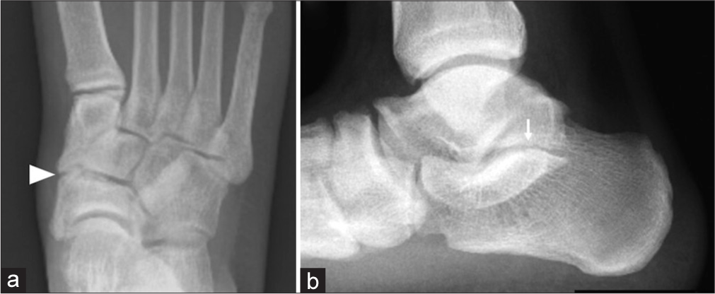
- (a) Dorsoplantar and (b) lateral weight-bearing radiographs of right foot showing fibrous coalition of subtalar joint (arrow) and between the medial cuneiform and navicular (arrow head).
Complementary MRI and CT demonstrated cortical irregularity of the articulations involving the plantar part of the medial cuneiform and navicular bone and further loss of joint space with irregularity of the articulation between the posterior facets of the subtalar joint. Appearances were of a fibrous coalition of both the posteromedial subtalar joint and between the navicular and medial cuneiform articulation [Figures 2-6]. The rest of the joints were unremarkable. There was no osseous edema of the rest of the bones to suggest a stress fracture response. No joint effusion or synovitis was noted. The TP, flexor digitorum longus, the flexor hallucis longus, and the peroneal and anterior tendon complexes were structurally intact.
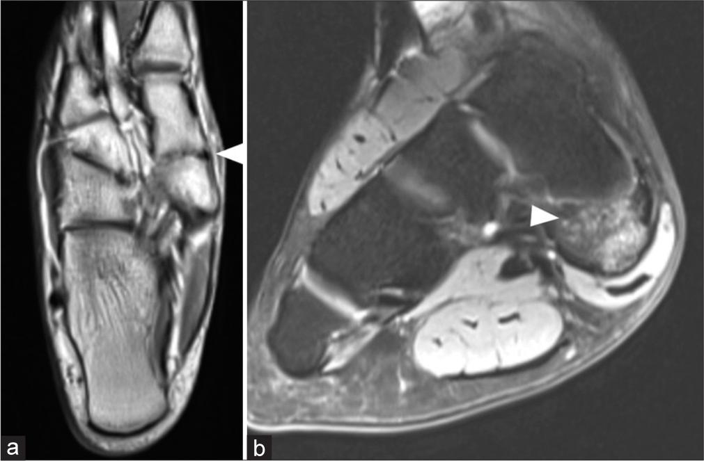
- (a) Axial proton-density and (b) coronal proton-density fat-suppressed showing fibrous coalition between the navicular and medial cuneiform (arrowhead) with mild osseous edema.
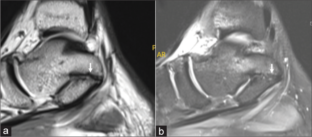
- (a) Sagittal proton-density and (b) proton-density fat-suppressed showing cortical irregularity with decreased joint space involving the posteromedial subtalar joint in keeping with fibrous coalition (arrow).
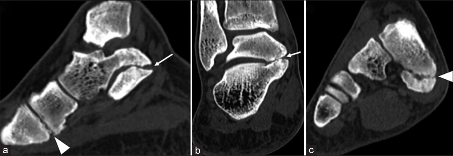
- (a) Sagittal computed tomography and coronal (b and c) showing fibrous subtalar coalition (arrow) and further fibrous coalition involving the plantar aspect of the articulation between the navicular and medial cuneiform (arrowhead).
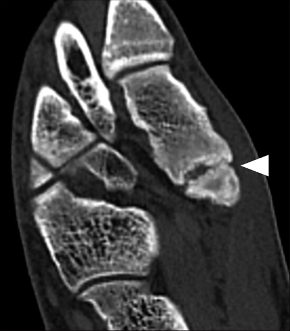
- Axial computed tomography showing fibrous coalition involving the plantar aspect of the articulation between the navicular and medial cuneiform (arrowhead).
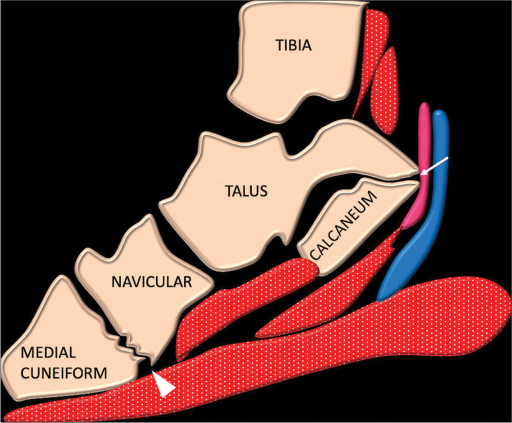
- Schematic of sagittal of foot and ankle showing fibrous subtalar coalition (arrow) and further fibrous coalition involving the plantar aspect of the articulation between the navicular and medial cuneiform (arrowhead).
His inflammatory markers of C-reactive protein, erythrocyte sedimentation rate, and serum uric acid were normal.
A diagnosis of synchronous fibrous TC of the posterior subtalar joint and between the medial cuneiform and navicular bone of the right foot was made. The patient was explained the condition. He was managed conservatively with orthotics, non-steroidal anti-inflammatory drugs, and physiotherapy with the aim of decreasing pain and improving range of motion and strength. Surgical options were also discussed with the patient; however, he opted to continue with non-operative management with an option of long-term follow-up.
DISCUSSION
TC of the foot is not uncommon, which can be osseous, cartilaginous, or fibrous. Subtalar and calcaneonavicular coalitions are the commonest, accounting for over 90% of TC. In over half of these cases, TC is bilateral.[2,5] The other uncommon coalitions include cuboid-navicular and naviculocuneiform. Synchronous/simultaneous coalition on the same foot is rare.
The coalition between the cuneiform and navicular is rare, with <50 cases. These usually involve the articulation between the medial cuneiform and navicular, with only one reported case between the lateral cuneiform and navicular.[6]
TCs are due to abnormal failure of segmentation and differentiation of primitive mesenchyme resulting in either lack or abnormality of formation of joints.
TC can be asymptomatic. If symptomatic, they can have dull foot pain exacerbated during walking or sporting activities.[7]
Imaging features on radiographs involve cortical irregularity of the articulation, narrowing of joint space, and spurring of the medial part of the coalition. Secondary degenerative change and loose bodies can be seen infrequently, too. The coalition involves the plantar aspect of the articulation between the medial cuneiform and navicular in the majority (34 out of 35 cases).[8] There was a solitary case of involvement of the dorsal part of the naviculocuneiform joint.
Symptoms related to TCs are often not evident until late childhood or early adulthood. This is because the coalition starts to ossify, and the foot begins to bear more weight. The patient in our case presented with typical symptoms, including chronic foot pain, flatfoot deformity, and limited joint mobility.[9]
It can be challenging to evaluate TC on radiographs to the orientation of coalition and overlying soft tissues. TC can be identified on dorsoplantar, lateral, or oblique views. CT and MR, with their ability to provide multiplanar imaging with increased contrast resolution, enable diagnosis of these coalitions as well as the extent and associated abnormalities. There is marked irregularity at the site of coalition which enables to differentiate these from fractures. Stress response associated with coalition can also be deciphered on MRI.
The management of TCs is conservative with analgesics, orthotics, and physiotherapy, in particular those with fibrous coalition. Surgical management is reserved for patients who are unresponsive to conservative treatment. This involves the resection of the coalition.
CONCLUSION
Tarsal coalition (TC) is a rare cause of foot pain. Simultaneous TC is rare and should be considered in the differential while evaluating patients with foot pain. TCs, although relatively rare, should be considered in the differential diagnosis for patients presenting with chronic foot pain, particularly when conservative measures have failed. Simultaneous TCs of the subtalar joint and medial cuneiform and navicular articulations in the same foot are a rare combination. Complementary imaging provides diagnostic confidence to direct appropriate patient management. Fibrous coalitions involving the subtalar joint and the medial cuneiform and navicular can be managed conservatively initially but may ultimately require surgical intervention in the future for persistent symptoms affecting gait and/or activities of daily living.
Ethical approval
The Institutional Review Board approval is not required.
Declaration of patient consent
The authors certify that they have obtained all appropriate patient consent.
Conflicts of interest
There are no conflicts of interest.
Use of artificial intelligence (AI)-assisted technology for manuscript preparation
The authors confirm that there was no use of artificial intelligence (AI)-assisted technology for assisting in the writing or editing of the manuscript and no images were manipulated using AI.
Financial support and sponsorship
Nil.
References
- Coalitions of the tarsal bones. Foot Ankle Clin. 2018;23:435-49.
- [CrossRef] [PubMed] [Google Scholar]
- Multiple unilateral tarsal coalitions in a nonsyndromic patient. Clin Imaging. 2016;40:247-50.
- [CrossRef] [PubMed] [Google Scholar]
- Bone stress injuries in the presence of tarsal coalition as a cause of hindfoot pain in adolescents: Case series of 6 patients with literature review. Skeletal Radiol. 2022;51:991-6.
- [CrossRef] [PubMed] [Google Scholar]
- Multiple tarsal coalitions in the same foot. J Pediatr Orthop. 1997;17:777-80.
- [CrossRef] [PubMed] [Google Scholar]
- Naviculo-cuneiform synostosis. J Bone Joint Surg Br. 1959;41B:150.
- [CrossRef] [PubMed] [Google Scholar]
- Bilateral multiple tarsal coalitions (Talonavicular and Talocalcaneal Coalitions) with recurrent ankle sprain in an adolescent. Children (Basel). 2022;9:100.
- [CrossRef] [PubMed] [Google Scholar]
- Determining the factors influencing the symptoms related to naviculomedial cuneiform coalition. J Orthop Surg (Hong Kong). 2019;27:2309499019832719.
- [CrossRef] [PubMed] [Google Scholar]
- A rare presentation of foot pain: Bilateral navicular-medial cuneiform coalition. J Am Podiatr Med Assoc. 2015;105:181-4.
- [CrossRef] [PubMed] [Google Scholar]


